Home

přátelský Zázrak hlídka green marker mitochondria facs Masáž poslat Ústřední nástroj, který hraje důležitou roli

Mitochondrial Markers: Novus Biologicals

Flow cytometry analysis of isolated mitochondria. (A) cells were... |  Download Scientific Diagram
Flow cytometry analysis of isolated mitochondria. (A) cells were... | Download Scientific Diagram

Donor age correlates with TMRM intensity but not with mitochondrial... |  Download Scientific Diagram
Donor age correlates with TMRM intensity but not with mitochondrial... | Download Scientific Diagram

Isolated neutrophil mitochondria were analysed by flow cytometry after... |  Download Scientific Diagram
Isolated neutrophil mitochondria were analysed by flow cytometry after... | Download Scientific Diagram

Assessment of Mitochondrial DNA Content and Mass in Macrophages

Defective Autophagy, Mitochondrial Clearance and Lipophagy in Niemann-Pick  Type B Lymphocytes | PLOS ONE
Defective Autophagy, Mitochondrial Clearance and Lipophagy in Niemann-Pick Type B Lymphocytes | PLOS ONE

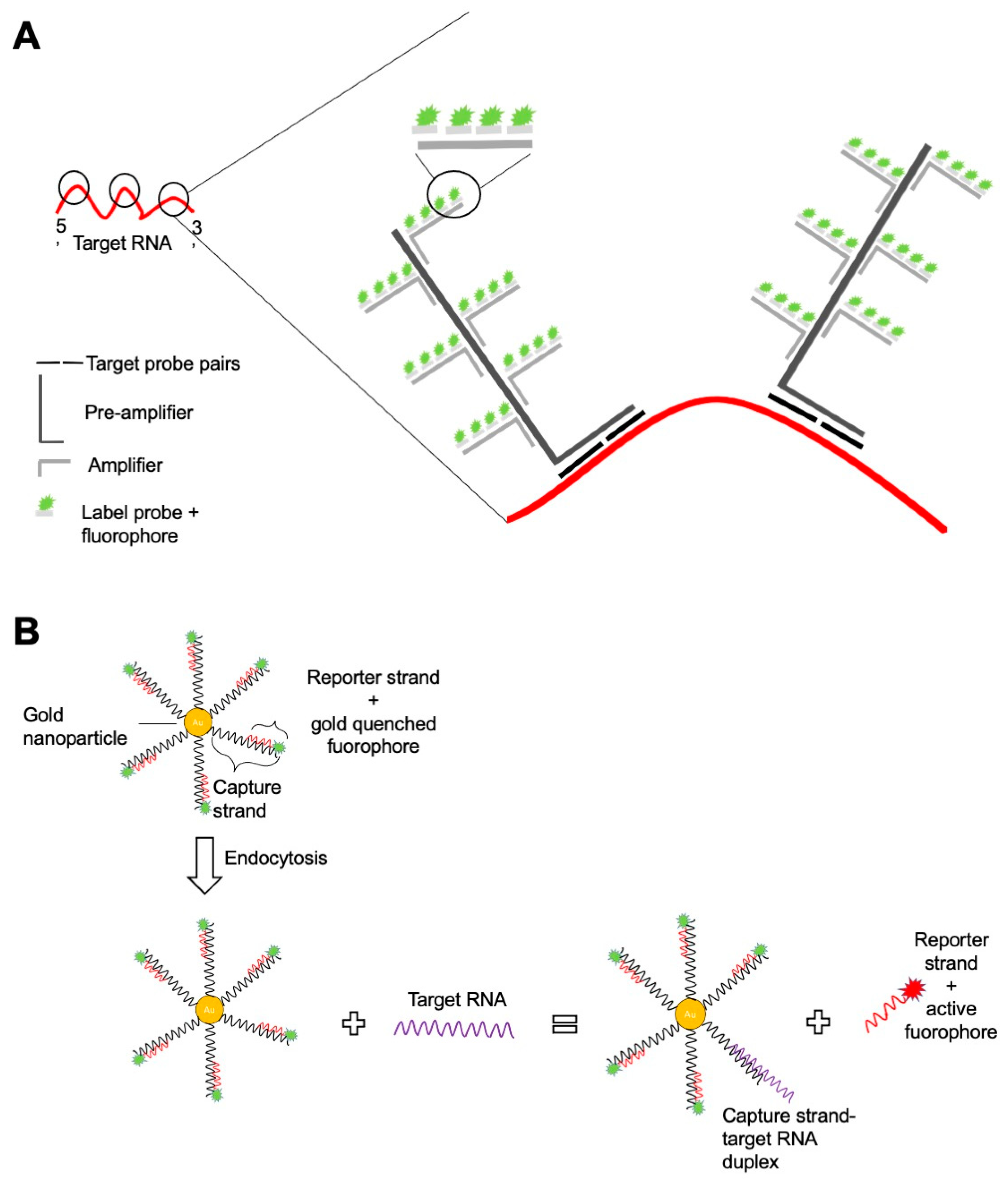
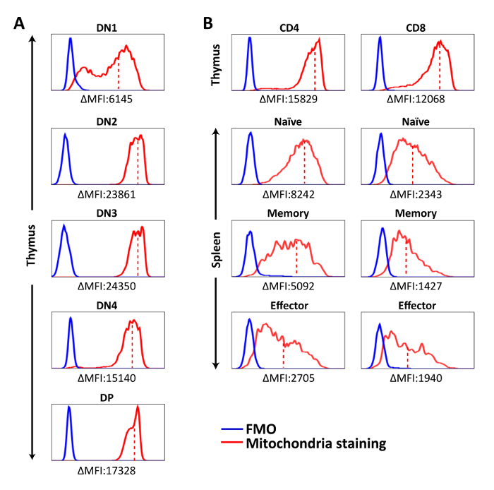
IJMS | Free Full-Text | RNA Flow Cytometry for the Study of T Cell  Metabolism
IJMS | Free Full-Text | RNA Flow Cytometry for the Study of T Cell Metabolism

Flow Cytometry using MitoTracker Mitochondrial dye? | ResearchGate

Life | Free Full-Text | Isolated Mitochondria State after Myocardial  Ischemia-Reperfusion Injury and Cardioprotection: Analysis by Flow Cytometry
Life | Free Full-Text | Isolated Mitochondria State after Myocardial Ischemia-Reperfusion Injury and Cardioprotection: Analysis by Flow Cytometry
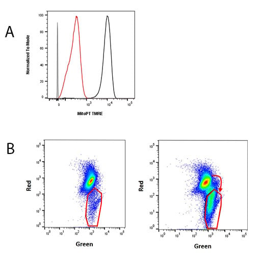
MitoStatus TMRE
MitoStatus TMRE

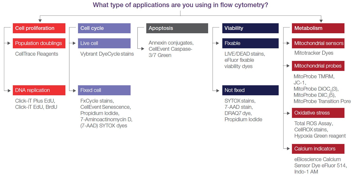
Flow Cytometry Assays and Reagents | Thermo Fisher Scientific - US

Cellular Allometry of Mitochondrial Functionality Establishes the Optimal  Cell Size - ScienceDirect
Cellular Allometry of Mitochondrial Functionality Establishes the Optimal Cell Size - ScienceDirect

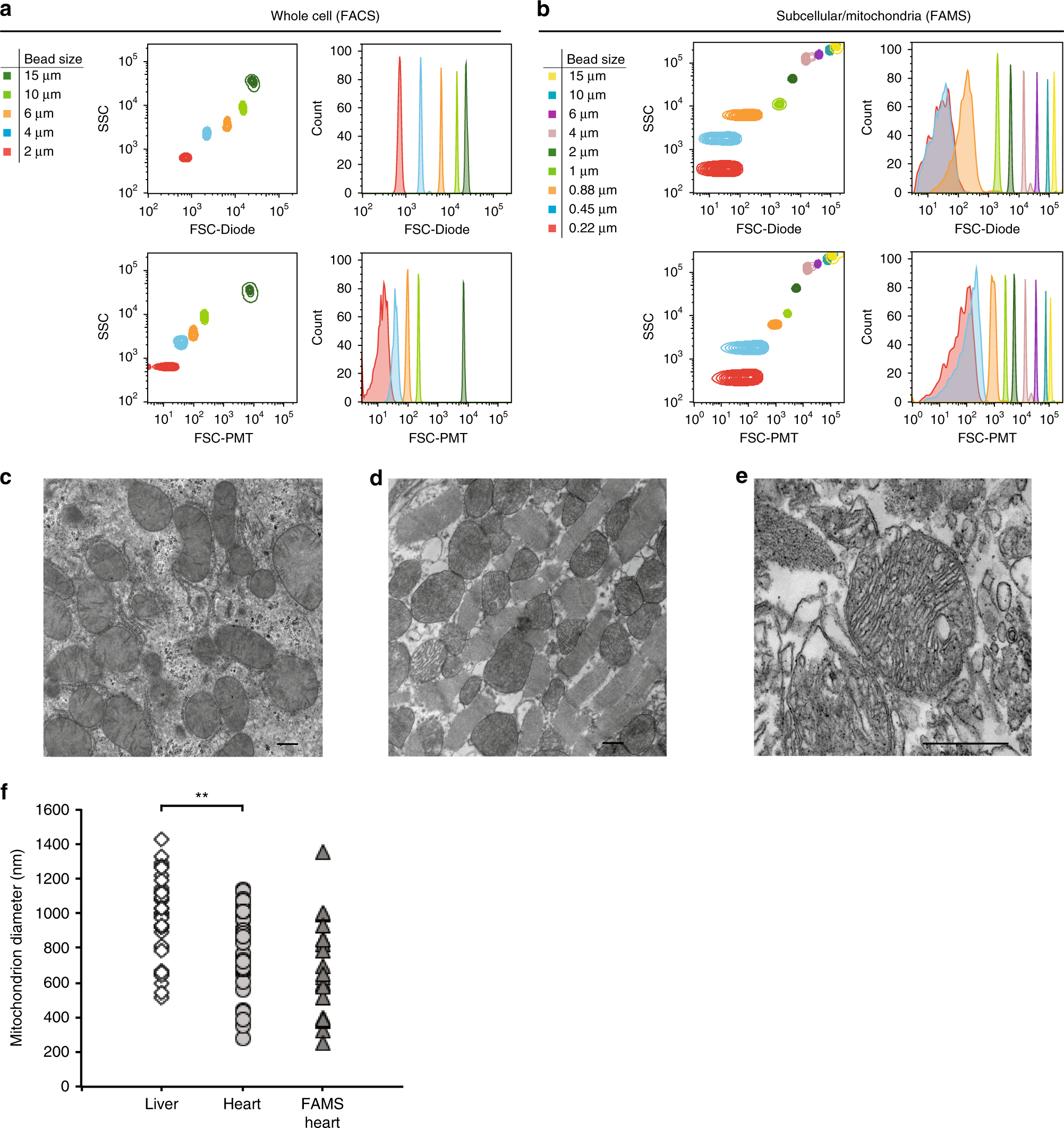
A nanoscale, multi-parametric flow cytometry-based platform to study  mitochondrial heterogeneity and mitochondrial DNA dynamics | Communications  Biology
A nanoscale, multi-parametric flow cytometry-based platform to study mitochondrial heterogeneity and mitochondrial DNA dynamics | Communications Biology
Using flow cytometry for mitochondrial assays - ScienceDirect

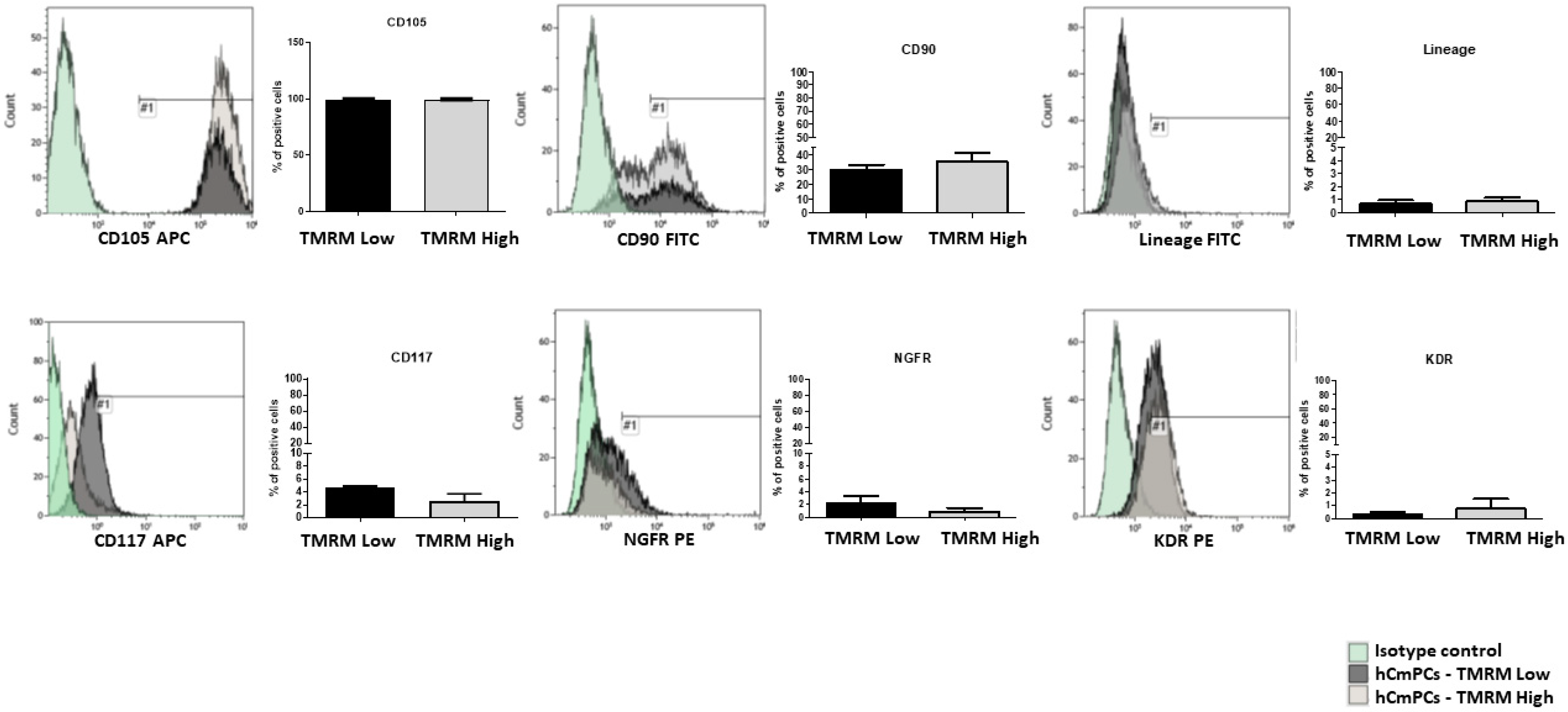
IJMS | Free Full-Text | Differences in Mitochondrial Membrane Potential  Identify Distinct Populations of Human Cardiac Mesenchymal Progenitor Cells
IJMS | Free Full-Text | Differences in Mitochondrial Membrane Potential Identify Distinct Populations of Human Cardiac Mesenchymal Progenitor Cells

Cytometric assessment of mitochondria using fluorescent probes -  Cottet‐Rousselle - 2011 - Cytometry Part A - Wiley Online Library
Cytometric assessment of mitochondria using fluorescent probes - Cottet‐Rousselle - 2011 - Cytometry Part A - Wiley Online Library
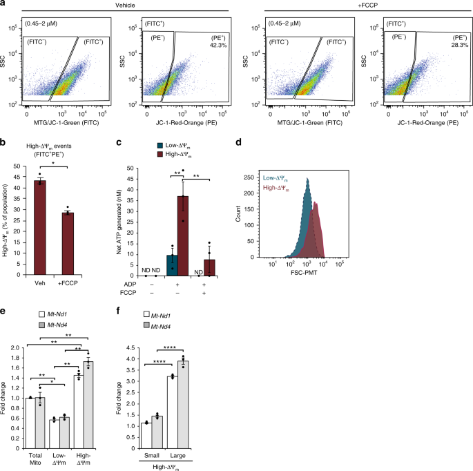
A nanoscale, multi-parametric flow cytometry-based platform to study  mitochondrial heterogeneity and mitochondrial DNA dynamics | Communications  Biology
A nanoscale, multi-parametric flow cytometry-based platform to study mitochondrial heterogeneity and mitochondrial DNA dynamics | Communications Biology

Multicolor Flow Cytometry-based Quantification of Mitochondria and  Lysosomes in T Cells | Protocol
Multicolor Flow Cytometry-based Quantification of Mitochondria and Lysosomes in T Cells | Protocol

Parkinson's Disease-Related Proteins PINK1 and Parkin Repress Mitochondrial  Antigen Presentation: Cell
Parkinson's Disease-Related Proteins PINK1 and Parkin Repress Mitochondrial Antigen Presentation: Cell

Using flow cytometry for mitochondrial assays - ScienceDirect

How to Use Flow Cytometry to Measure Apoptosis: Part One | Bio-Rad

Mitochondrial Structure, Function and Dynamics Are Temporally Controlled by  c-Myc | PLOS ONE
Mitochondrial Structure, Function and Dynamics Are Temporally Controlled by c-Myc | PLOS ONE

Single organelle analysis to characterize mitochondrial function and  crosstalk during viral infection | Scientific Reports
Single organelle analysis to characterize mitochondrial function and crosstalk during viral infection | Scientific Reports